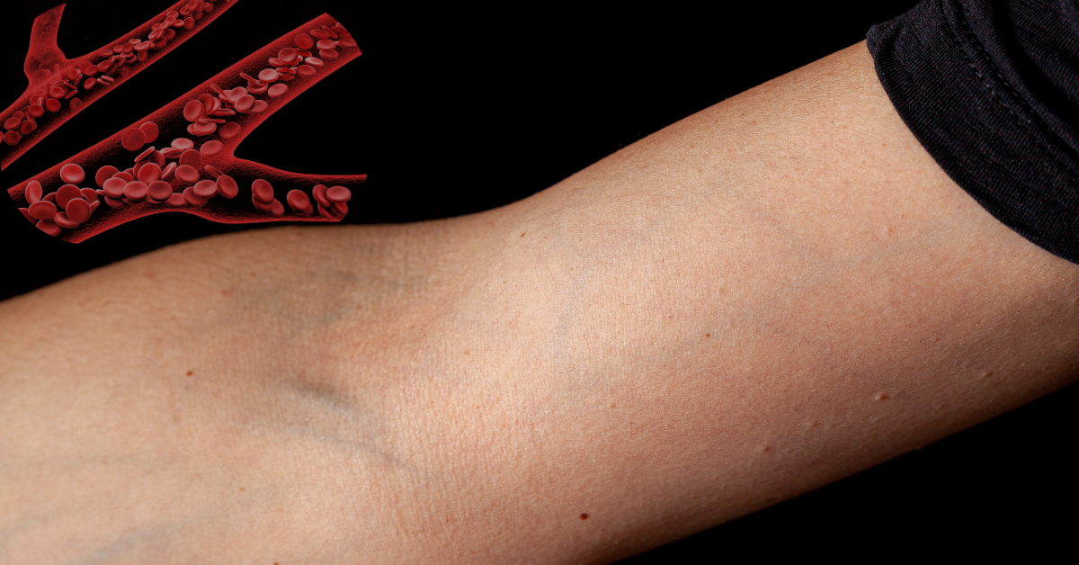
With its various functions ranging from venous return to thermoregulation, the cephalic vein is an essential part of the upper limb’s venous system. Cephalic Vein: Key Insights-This comprehensive guide examines the cephalic vein’s anatomy, physiology, clinical relevance, and related disorders to provide readers with a thorough grasp of the vein’s significance in medicine.
1. Beginning and Course
The dorsal venous network of the hand, more especially the radial side, is where the cephalic vein begins. It starts as a portion of the upper limb’s superficial venous system, where smaller veins from the thumb and the radial side of the hand merge. The vein travels in a lateral direction along the forearm, running next to the brachioradialis muscle. It is reasonably straightforward to palpate and access because of this muscle’s prominence and the vein’s superficial route.
The cephalic vein climbs over the lateral portion of the forearm and upper arm, eventually joining the axillary vein at the shoulder, close to the acromion process and deltoid muscle. Because the vein is shallow and easier to reach than deeper veins, this route is essential for doctors doing operations like venipuncture and intravenous cannulation. It also lowers the risk of complications.
2. Interaction with other frameworks-Cephalic Vein: Key Insights
The brachial artery and the biceps brachii muscle have superficial connections to the cephalic vein. Venipuncture benefits from the brachial artery and biceps brachii’s deeper placement than the vein. The proximity of the acromion process and deltoid muscle provides additional reference points during catheter insertion or surgical procedures.
For instance, because of its convenience and low risk of major artery damage, doctors frequently favor the cephalic vein’s accessibility for procedures like the implantation of a central venous catheter or emergency treatments.
3. Various-Cephalic Vein: Key Insights
The cephalic vein has a variety of anatomical variants. Changes in its diameter, course, or the existence of new tributaries are examples of variations. Some people may have many branches or a split cephalic vein, which makes it more difficult to visually identify and use for medical purposes.
In some cases, the cephalic vein exhibits several tributaries from the forearm or joins the subclavian vein directly. These differences may impact the methods used in procedures, necessitating careful planning and imaging.

Anatomical Structure
1. Return of Venous Blood
The cephalic vein is an essential part of deoxygenated blood’s venous return from the upper limb to the heart. It is essential for preserving systemic venous pressure and facilitating blood flow back to the right atrium. This role maintains effective cardiovascular function and supports overall circulatory homeostasis.
When chronic venous insufficiency impairs efficient venous return, the cephalic vein plays a crucial role in venous return. The cephalic vein assists in this return and, as a result, helps control systemic blood pressure and volume, particularly in situations where there are large fluid shifts or venous pressure fluctuations.
2. Controlling body temperature-Cephalic Vein: Key Insights
In addition to venous return, the cephalic vein aids in thermoregulation. It contributes to maintaining core body temperature and dissipating metabolic heat by adjusting blood flow. This function is critical when engaging in physical activity or under different environmental circumstances.
When exercising vigorously, for example, the cephalic vein helps the body cool by facilitating the movement of warmer blood from the core to the periphery, where heat loss may happen more easily. The body’s temperature stays within a healthy range even during stressful situations because of the cephalic vein’s effective thermoregulation, which helps avoid diseases like hyperthermia.
3. Significance in Clinical Practice
Taking care of illnesses connected to venous insufficiency, like chronic venous diseases or issues after placing a catheter, requires a thorough knowledge of how the cephalic vein works physiologically. Due to the vein’s dual function in venous return and thermoregulation, it is important to be precise in medical treatment. This is because the vein can be used for a wide range of therapeutic procedures, from simple venipuncture to complex vascular operations.
Use in Clinical Settings
1. Venipuncture and cannulation
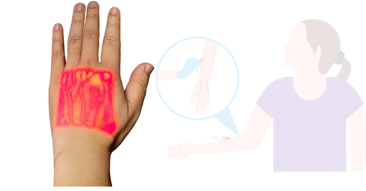
The cephalic vein is a favorable site for intravenous cannulation and venipuncture due to its superficial placement. Its visibility and accessibility minimize risks such as accidental artery puncture or nerve damage, making it the perfect option for routine blood draws and intravenous treatments.
Emergency scenarios frequently utilize the cephalic vein to provide quick intravenous access, enabling immediate treatment. In order to reduce risks and guarantee successful venous access, proper diagnosis and technique are essential. Methods such as ultrasonography guidance can improve cannulation precision and reduce problems.
2. Dialysis Entry-Cephalic Vein: Key Insights
People who require chronic dialysis frequently use the cephalic vein to establish arteriovenous fistulas. In order to improve blood flow for hemodialysis, this surgical treatment entails joining the cephalic vein to an adjacent artery. Such operations depend on exacting surgical skill and cautious postoperative care to avoid problems like thrombosis or infection.
A well-constructed arteriovenous fistula provides dependable access for repeated dialysis sessions to treat chronic renal failure.
3. A Look at Surgical Considerations
To prevent unintentional harm during upper-limb procedures, it is essential to understand the anatomy of the cephalic vein. In order to avoid problems like thrombosis or excessive bleeding, surgeons must avoid this vein. Ultrasound and CT scans are examples of preoperative imaging methods that can help with planning and risk reduction.
Knowing the exact position of the cephalic vein, for instance, can help ensure the best possible surgical results and prevent unintentional harm during orthopedic or reconstructive procedures.

Imaging and diagnostic methods
1. Computed sonography
An essential imaging modality for assessing the cephalic vein is ultrasound. In addition to helping to guide cannulation techniques and detect thrombi or structural anomalies, it offers real-time viewing of the vein’s patency. When it comes to evaluating blood flow dynamics, identifying ailments such as venous insufficiency or stenosis, and planning remedies, Doppler ultrasonography is very helpful.
2. Studies with Contrast-Enhanced
In more complicated situations, we may use contrast-enhanced imaging methods like MRIs and CT venography. These cutting-edge techniques make it easier to diagnose venous anomalies or determine disease severity by providing precise images of the cephalic vein and its surrounding tissues.
For example, you can use contrast-enhanced CT venography to map out the venous architecture before surgical operations or to assess suspected venous thrombosis.
Pathological States
1. Diphtheria
Extended intravenous catheterization frequently links to thrombophilia, an inflammatory disorder. Pain, erythema, and swelling along the cephalic vein are the symptoms. To treat symptoms and prevent further problems, management typically includes applying heat, using anti-inflammatory drugs, and, if required, removing the catheter. In extreme situations, thrombolytic treatment may be considered to remove the clot and restore normal venous flow.
2. Swollen veins
In the cephalic vein, varicose veins are less prevalent than deeper veins, although they can still happen. Treatment is necessary to relieve symptoms and avoid the consequences of varicose cephalic veins, which can manifest as twisted, engorged veins. One option for management is scleotherapy, which involves injecting a sclerosing agent to collapse the vein. In more severe situations, surgery may be necessary.
3. Injuries
Significant bleeding or difficulties can result from trauma to the cephalic vein, which can happen as a result of external forces or surgical operations, can cause significant bleeding or difficulties. To manage any bleeding and reduce unfavorable consequences, timely identification and intervention are essential. Direct pressure to stop bleeding may be part of the immediate treatment, and if surgery is required, that will come next.
In conclusion
The clinical significance and strategic anatomical placement of the cephalic vein highlight its role in the venous system. A comprehensive understanding of its anatomy, physiological functioning, and potential diseases enhances medical practice and improves patient care. In both health and sickness, the cephalic vein is essential for regular clinical treatments as well as intricate surgical techniques. We need further research and comprehension in this area to improve patient outcomes and advance therapeutic procedures.
Clinicians can improve procedure results, better treat a variety of disorders, and provide better overall patient care by developing a greater understanding of the cephalic vein.
FAQ:
What is the cephalic vein?
The cephalic vein is a prominent vein in the upper limb that originates from the dorsal venous network of the hand. It travels along the lateral aspect of the forearm and arm, eventually joining the axillary vein near the shoulder. It plays a crucial role in venous return from the upper limb and is commonly used for venipuncture and intravenous access
Where is the cephalic vein located?
The cephalic vein runs superficially along the lateral side of the forearm and upper arm. It is positioned just beneath the skin, making it easily accessible for clinical procedures such as blood draws or catheter placements
What are common uses of the cephalic vein in medical procedures?
The cephalic vein is frequently used for venipuncture, intravenous cannulation, and creating arteriovenous fistulas for dialysis. Its superficial location makes it a preferred site for these procedures due to ease of access
What are potential complications associated with the cephalic vein?
Complications related to the cephalic vein include thrombophlebitis, which is inflammation and clotting in the vein, and varicose veins. Trauma to the cephalic vein can also lead to significant bleeding or complications during medical procedures
How can imaging techniques help in assessing the cephalic vein?
Ultrasound imaging is commonly used to visualize the cephalic vein, aiding in procedures like cannulation and detecting conditions such as thrombosis. For more detailed views, contrast-enhanced CT venography or MRI can be employed to assess the vein’s structure and associated pathologies


